Smooth Muscle Diagram / Smooth muscle determines the flow of blood in the arteries.. This smooth muscle can be found surrounding the walls of the blood vessels, the bronchioles in the lungs, and the sphincter muscles used in the gi tract.the gi tract, which is tubular by design, also houses longitudinal muscles in addition to the smooth. Smooth muscle contraction requires both myosin activation and actin cytoskeletal remodeling. It is the pen diagram of skeletal, smooth and cardiac muscle for class 10, 11 and 12. Draw a diagram of smooth muscle fibre and label any three parts. It constitutes much of the musculature of
It also occurs in the spleen (capsule and trabeculae), eye (iris and ciliary body), skin (arrector pili muscles of hairs. Smooth muscles are structurally the simplest of all muscles types. Arteries have thick walls due to smooth muscle cells, which help them carry blood away from the heart to every part of. • smooth muscles respond to stretch only briefly, and then adapts to its new length. Draw a diagram of smooth muscle fibre and label any three parts.
Draw A Labelled Diagram Of Smooth Muscle Explain Brainly In from hi-static.z-dn.net Molecular biology of the cell. Smooth muscle tissue, unlike striated muscle, contracts slowly and automatically. In this video i have shown the simplest way of drawing muscle drawing. These cells have fibers of actin and myosin which run through the cell and are supported by a framework of other proteins. Smooth muscle is found in the walls of hollow organs, including the stomach, intestines, bladder. The contractile property of muscles is used effectively to bring about a movement. Smooth muscle, muscle that shows no cross stripes under microscopic magnification. Smooth muscle muscle tissue in which the contractile fibrils are not highly ordered, occurring in the gut and other internal organs and not under voluntary control.
They are characterized by an assembly of short, narrow cells with differing properties.
• smooth muscles respond to stretch only briefly, and then adapts to its new length • the new length however, retains its original _____ seconds or minutes after it has been elongated or shortened (e.g. The posterior wall of the trachea not covered by cartilage is composed of connective tissue and smooth muscle. Smooth muscle is widely distributed in the body. The muscle tissue (patrick steele) These cells have fibers of actin and myosin which run through the cell and are supported by a framework of other proteins. Smooth muscles exhibits a phenomenon called _____ in which: Smooth muscle determines the flow of blood in the arteries. Related posts of smooth muscle diagram human anatomy lymphatic system. It constitutes much of the musculature of Smooth muscle contraction requires both myosin activation and actin cytoskeletal remodeling. Draw a diagram of smooth muscle fibre and label any three parts. Human muscles · august 4, 2020. Compared with skeletal muscles, smooth muscle cells contract and relax slowly, and they can create and maintain tension for long periods of time.
Smooth muscle makes up the walls of hollow organs, respiratory passageways, and blood vessels. Smooth muscle diagram, find out more about smooth muscle diagram. Smooth muscle is a type of muscle tissue which is used by various systems to apply pressure to vessels and organs. This smooth muscle can be found surrounding the walls of the blood vessels, the bronchioles in the lungs, and the sphincter muscles used in the gi tract.the gi tract, which is tubular by design, also houses longitudinal muscles in addition to the smooth. Transcribed image text from this question.
Proposed Model For Mechanism Of Smooth Muscle Plasticity Illustrating Download Scientific Diagram from www.researchgate.net It is found in the walls of ducts and blood and lymphatic vessels, as well as in the walls of the digestive, respiratory and urogenital tracts. The posterior wall of the trachea not covered by cartilage is composed of connective tissue and smooth muscle. This diagram shows a few of the cells that can be seen in the stained section below. Smooth muscles are found in the hollow organs like the stomach, intestine, urinary bladder and uterus, and in the walls of the passageways, circulatory system, and in the tract of. Smooth muscle is composed of sheets or strands of smooth muscle cells. The muscle tissue (patrick steele) The calcium is the cause of protein to detach from the actin and myosin fastly binds with the opening of actin. It constitutes much of the musculature of
The contractile property of muscles is used effectively to bring about a movement.
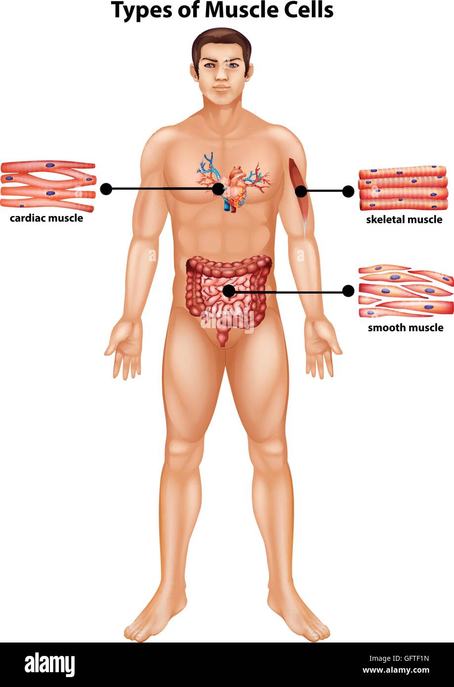
The three types present are: It constitutes much of the musculature of The cells stick together and are connected by specialised cell junctions, called gap junctions. Arteries have thick walls due to smooth muscle cells, which help them carry blood away from the heart to every part of. Transcribed image text from this question. It is layered in a distinctive pattern of circular layers. Vascular smooth muscle is innervated primarily by the sympathetic nervous system through adrenergic receptors (adrenoceptors). Compared with skeletal muscles, smooth muscle cells contract and relax slowly, and they can create and maintain tension for long periods of time. Smooth muscle contracts under certain stimuli as atp is freed. It is the pen diagram of skeletal, smooth and cardiac muscle for class 10, 11 and 12. Smooth muscle is made up of cells that contain a single central nucleus. The cells are spindle shaped, and the nucleus is central. • smooth muscles respond to stretch only briefly, and then adapts to its new length • the new length however, retains its original _____ seconds or minutes after it has been elongated or shortened (e.g.
Drawing exercise sectional smooth muscle tissue labeled diagram | world of reference. It is the pen diagram of skeletal, smooth and cardiac muscle for class 10, 11 and 12. The three types present are: It is found in the walls of ducts and blood and lymphatic vessels, as well as in the walls of the digestive, respiratory and urogenital tracts. Smooth muscle anatomy smooth muscle tissue is also known as visceral muscle tissue.
Smooth Muscle High Resolution Stock Photography And Images Alamy from c8.alamy.com Smooth muscle is made up of cells that contain a single central nucleus. They are characterized by an assembly of short, narrow cells with differing properties. Smooth muscle (textus muscularis levis) smooth muscle is a type of tissue found in the walls of hollow organs, such as the intestines, uterus and stomach. Diagram of a small artery in cross section. The calcium is the cause of protein to detach from the actin and myosin fastly binds with the opening of actin. The cells are spindle shaped, and the nucleus is central. In this video i have shown the simplest way of drawing muscle drawing. Smooth muscle anatomy smooth muscle tissue is also known as visceral muscle tissue.
The smooth muscles perform the functions in the contrast of other types of muscles.
Smooth muscle contraction requires both myosin activation and actin cytoskeletal remodeling. Molecular biology of the cell. The muscle tissue (patrick steele) Smooth muscles are found in the hollow organs like the stomach, intestine, urinary bladder and uterus, and in the walls of the passageways, circulatory system, and in the tract of. • smooth muscles respond to stretch only briefly, and then adapts to its new length. Smooth muscle, muscle that shows no cross stripes under microscopic magnification. Smooth muscle anatomy smooth muscle tissue is also known as visceral muscle tissue. In arteries, smooth muscle movements maintain the arteries' diameter. Its wavelike movements propel things through the bodily system, such as food through. Smooth muscle diagram, find out more about smooth muscle diagram. The contractile property of muscles is used effectively to bring about a movement. The posterior wall of the trachea not covered by cartilage is composed of connective tissue and smooth muscle. This diagram shows a few of the cells that can be seen in the stained section below.


0 Komentar